The images provided offer a striking and unfiltered look into severe dermatological conditions that go far beyond the common pimple. They depict chronic, progressive skin disorders that can have profound physical and psychological effects on those who live with them.
The primary condition shown in the first image is an advanced case of Rhinophyma, accompanied by numerous giant comedones (large blackheads). The second image further illustrates different types of significant skin impactions undergoing professional extraction.
This article will explore the science behind these conditions, debunk common myths, and detail the medical treatments available.
Section 1: Understanding Rhinophyma
The first image shows a nose that has undergone a dramatic transformation. The skin is thickened, irregular, and bulbous. This condition is known as Rhinophyma (pronounced ry-no-fy-ma).
What Is It?
Rhinophyma is classified as a severe, late-stage subtype of rosacea, specifically phymatous rosacea. While common rosacea typically presents as facial redness, visible blood vessels, and small bumps, phymatous rosacea involves actual structural changes to the skin.
The Pathology of Growth
The deformity seen in the photo is caused by a slow, progressive overgrowth (hypertrophy) of two key components of the skin:
-
Sebaceous (Oil) Glands: The nose has a high concentration of oil glands. In Rhinophyma, these glands enlarge significantly and become hyperactive, producing excessive amounts of sebum (skin oil).
-
Connective Tissue: The surrounding tissue and collagen thicken, leading to fibrosis (scar-like tissue formation).
This combined overgrowth causes the skin to expand, creating the lumpy, lobulated, and enlarged appearance characteristic of the condition.
Debunking the “Drinker’s Nose” Myth
For generations, a bulbous, red nose was unfairly and incorrectly associated with heavy alcohol consumption, leading to stigmatizing terms like “gin blossom” or “whiskey nose.”
This is a medical myth.
While alcohol acts as a vasodilator, causing temporary flushing that can exaggerate the redness of rosacea, it does not cause Rhinophyma. The condition is primarily driven by genetics, age, and hormonal factors. It is overwhelmingly more common in men than women and typically develops between the ages of 50 and 70. People who have never consumed alcohol can and do develop Rhinophyma.
Section 2: The Anatomy of Giant Comedones
A dominant feature in both images is the presence of large, dark, crater-like pores. These are comedones, the medical term for clogged pores.
Why Are They So Big and Black?
The “black” in a blackhead is not dirt. It is a plug formed by a mixture of excess sebum (oil) and dead skin cells (keratin) that gets trapped in the hair follicle. When this plug is open to the skin’s surface, the air causes the melanin in the dead skin cells to oxidize, turning it a dark brown or black color.
In the case of the Rhinophyma patient (Image 1), the massive size of these blackheads is a direct result of the condition. The enlarged, overactive oil glands pump out immense amounts of sebum. Simultaneously, the thickening, distorted skin structure stretches the pores, allowing huge plugs of debris to accumulate over time.
Dilated Pore of Winer
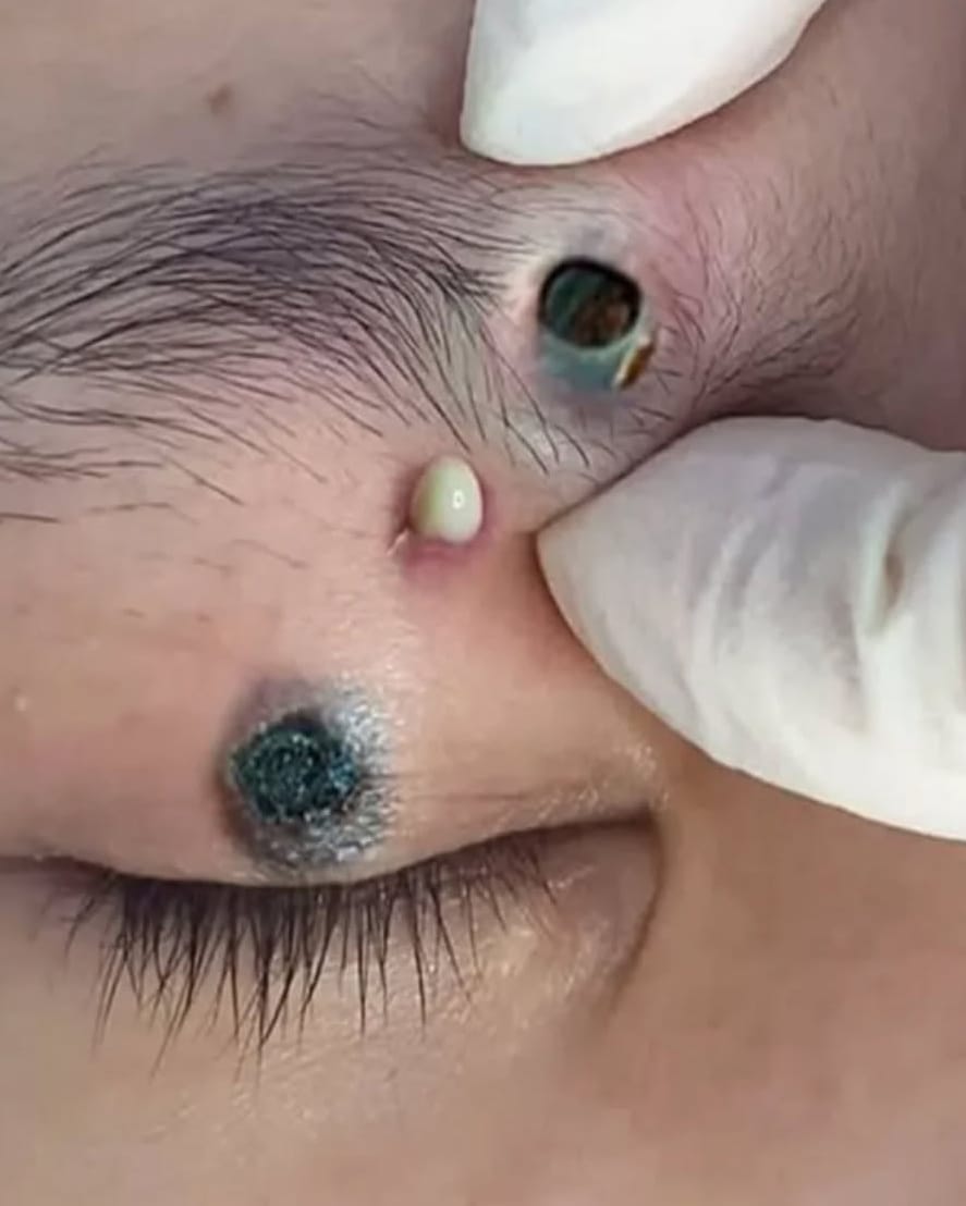
The second image shows a large, dark, distinct lesion near the eyebrow. This is likely a Dilated Pore of Winer. This is essentially a solitary, giant blackhead that has grown so large it has permanently dilated the pore structure, creating a benign, tumor-like lesion filled with dense keratin.
The same image also shows a gloved hand expressing a white lesion. This is a closed comedo (whitehead) or a small pustule, where the trapped oil and pus are not exposed to air and remain white or yellowish.
Section 3: Treatment and Restoration
The gloved hands visible in both images indicate that professional medical intervention is taking place. Treating these conditions requires more than over-the-counter creams; it often involves physical and surgical procedures.
1. Professional Extraction
For giant comedones and Dilated Pores of Winer, a dermatologist or skincare professional will perform manual extractions using sterile tools to clear the impacted debris. This is a crucial first step to reduce inflammation and prepare the skin for further treatment. It is highly discouraged to attempt this at home, as it can lead to severe infection and permanent scarring.
2. Treating Rhinophyma
The treatment for Rhinophyma aims to remove the excess tissue and reshape the nose. This is considered a reconstructive procedure and is often described by patients as life-changing.
-
Medical Management (Early Stages): In the very early stages of phymatous rosacea, oral isotretinoin (Accutane) may be prescribed to shrink the oil glands and slow progression. However, it cannot reverse existing fibrous tissue growth.
-
Surgical Resurfacing (Advanced Stages): For a nose like the one in the image, surgery is the gold standard. Common methods include:
-
CO2 Laser Ablation: Using a carbon dioxide laser to precisely vaporize layers of thickened skin, “sculpting” the nose back to a normal contour. This is often preferred for its precision and bloodless nature.
-
Electrosurgery: Using a heated wire loop to shave away excess tissue.
-
Scalpel Excision: Traditional surgical shaving of the bulbous tissue.
-
Conclusion
The conditions shown in these images are severe but treatable medical disorders. Rhinophyma can cause functional problems, such as nasal obstruction, in addition to significant psychosocial distress due to disfigurement. Modern dermatological surgery offers effective solutions, allowing patients to restore not just the appearance of their nose, but also their confidence and quality of life.